How Post-Inflammatory Hyperpigmentation Forms After Acne
Learn how post-inflammatory hyperpigmentation forms after acne breakouts and explore gentle topical ingredients like niacinamide to support even skin tone.
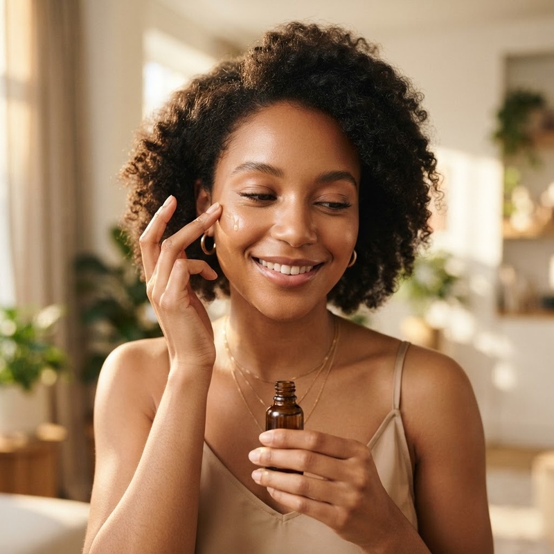
Navigating the lifecycle of an acne breakout can often feel like a multi-stage journey. For many individuals, once the active breakout subsides, the skin surface may not immediately return to its baseline appearance. Instead, flat brown, tan, dark, or pinkish marks frequently linger in the exact locations where blemishes once existed. This common occurrence can feel frustrating, especially when it seems as though skin clarification is taking longer than expected. However, understanding that these post-acne marks are a natural, protective biological response rather than permanent damage can offer clarity and peace of mind.
Navigating the lifecycle of an acne breakout can often feel like a multi-stage journey. For many individuals, once the active breakout subsides, the skin surface may not immediately return to its baseline appearance. Instead, flat brown, tan, dark, or pinkish marks frequently linger in the exact locations where blemishes once existed. This common occurrence can feel frustrating, especially when it seems as though skin clarification is taking longer than expected. However, understanding that these post-acne marks are a natural, protective biological response rather than permanent damage can offer clarity and peace of mind.
In clinical terms, these lingering marks are known as post-inflammatory hyperpigmentation (PIH) or post-inflammatory erythema (PIE). While they are often casually referred to as acne scars, true PIH is distinct from structural scarring because it does not involve permanent tissue loss or fibrous tissue overgrowth. Rather, PIH represents localized melanin accumulation triggered by the cascade of cutaneous inflammation. Research suggests that by understanding the cellular pathways that drive pigment formation, individuals can adopt thoughtful, barrier-supportive skincare habits. Gentle topical ingredients—such as niacinamide and azelaic acid—can support natural skin renewal processes and help diminish the appearance of lingering discoloration over time.
Understanding the Inflammatory Response in Acne-Prone Skin
To understand why dark spots form after acne heals, it helps to examine what occurs within the skin during an active blemish. Acne vulgaris begins deep within the pilosebaceous unit, which consists of a hair follicle and its associated sebaceous (oil) gland. When excess sebum combines with accumulated dead skin cells, microcomedones form, creating an oxygen-deprived environment. This localized environment encourages the growth of cutaneous bacteria, such as Cutibacterium acnes.
The Cellular Cascade of Cutaneous Blemishes
In response to bacterial proliferation and follicle distension, the innate immune system activates a precise cellular defense mechanism. White blood cells, including neutrophils and macrophages, migrate into the tissue surrounding the follicle. As these immune cells work to manage the localized disruption, they release chemical signaling molecules known as pro-inflammatory cytokines, chemokines, and reactive oxygen species (ROS). This cascade leads to localized redness, swelling, and warmth—the classic hallmark signs of cutaneous inflammation.
While this inflammatory cascade is essential for tissue defense and repair, the surrounding cellular architecture experiences collateral stimulation. Epidermal cells and underlying melanocytes (the specialized cells responsible for producing skin pigment) absorb these chemical distress signals, initiating biological changes that extend well past the resolution of the initial blemish.
Distinguishing PIH from Post-Inflammatory Erythema (PIE)
It is clinically valuable to distinguish post-inflammatory hyperpigmentation from post-inflammatory erythema, as their underlying biological causes differ. Post-inflammatory hyperpigmentation (PIH) arises from excess melanin production within the epidermal or dermal layers of the skin, typically appearing as tan, brown, or dark brown patches. PIH is particularly common in medium to darker skin phototypes (Fitzpatrick skin types III through VI), where melanocytes exhibit greater sensitivity and higher baseline activity levels.
Conversely, post-inflammatory erythema (PIE) stems from damaged, dilated, or inflamed microvascular capillaries located in the superficial dermis. PIE typically manifests as pink, red, or purplish discoloration and is more frequently observed in fair skin phototypes (Fitzpatrick types I and II). Understanding whether lingering discoloration is primarily driven by pigment accumulation (PIH) or vascular dilation (PIE) helps individuals select the most appropriate topical supportive ingredients for their skin routine.
The Biological Mechanism of Localized Melanin Overproduction
The core mechanism behind post-inflammatory hyperpigmentation centers on the complex interaction between inflammatory signaling molecules and epidermal melanocytes. Melanocytes are specialized dendritic cells situated along the basal layer of the epidermis, accounting for roughly 5% to 10% of the cells in the basal layer. Their primary biological function is synthesizing melanin, the pigment responsible for protecting skin tissue against ultraviolet radiation and oxidative stress.
Melanocyte Stimulation and Tyrosinase Activation
When an acne lesion induces tissue inflammation, inflammatory mediators—including prostaglandins (such as PGE2), leukotrienes (LTC4 and LTD4), histamine, thromboxane B2, and nitric oxide—are released into the surrounding extracellular matrix. These molecules bind to specific receptors located on the cell membrane of adjacent melanocytes.
This receptor binding triggers intracellular signaling pathways, notably upregulating the activity of tyrosinase, the rate-limiting enzyme involved in melanogenesis. Tyrosinase catalyzes the oxidation of the amino acid L-tyrosine into L-DOPA (dihydroxyphenylalanine), which subsequently undergoes further enzymatic conversions to synthesize eumelanin (brown/black pigment) or pheomelanin (yellow/red pigment). In the context of inflammatory stimulation, melanocytes increase their rate of melanin production, resulting in localized hyper-pigment deposition.
Melanosome Transfer and Epidermal Pigment Retention
Once synthesized, melanin pigment is packaged into membrane-bound intracellular vesicles known as melanosomes. Melanocytes utilize their dendritic extensions to transfer these pigment-laden melanosomes into neighboring epidermal keratinocytes. Each individual melanocyte maintains contact with approximately 36 keratinocytes, forming an epidermal melanin unit.
As keratinocytes receive these excess melanosomes, pigment accumulates above the cellular nuclei to protect DNA from light-induced damage. As keratinocytes naturally migrate upward through the epidermal layers during the skin's normal 28-to-40-day renewal cycle, the localized excess melanin moves toward the stratum corneum, creating a visible dark spot on the skin surface.
The depth of pigment deposition influences how discoloration resolves over time. In epidermal PIH, excess melanin remains confined within the epidermis and typically fades progressively as surface cells shed. In dermal PIH, inflammation disrupts the basement membrane separating the epidermis from the dermis, allowing melanin pigment to leak into the papillary dermis. Dermal melanophages (immune cells that engulf pigment) trap the melanin in deeper layers, resulting in discoloration that takes significantly longer to diminish.
Key Factors That Exacerbate Post-Acne Discoloration
While the initial inflammatory blemish triggers the onset of post-inflammatory hyperpigmentation, several external and internal factors can intensify melanin synthesis or delay natural skin recovery.
Ultraviolet Radiation and High-Energy Visible Light Exposure
Exposure to ultraviolet (UV) radiation—both UVA and UVB rays—is one of the most prominent external factors that exacerbates post-acne marks. UV light acts as a direct stimulant for melanogenesis. When newly healed or inflamed skin tissue is exposed to sunlight without protection, melanocytes respond by producing additional melanin to shield the tissue. This solar stimulation can darken existing spots and cause discoloration to persist for months longer than it otherwise would. Additionally, emerging research suggests high-energy visible (HEV) blue light from digital displays and sunlight may also contribute to prolonged hyperpigmentation in melanin-rich skin types.
Physical Picking and Aggressive Exfoliation
Mechanical trauma—such as squeezing, popping, or picking at acne blemishes—substantially intensifies the inflammatory response. Squeezing a blemish forces inflammatory debris deeper into the dermal tissue, disrupting the basement membrane and increasing the probability of dermal pigment entrapment. Similarly, using harsh physical scrubs or aggressive manual exfoliation tools on inflamed skin can create micro-tears, worsening localized irritation and triggering fresh pigment overproduction.
Hormonal Signals and Barrier Disruption
Systemic factors, including hormonal shifts, also influence melanocyte sensitivity. Estrogen and progesterone can modulate melanocortin receptor expression, rendering melanocytes more reactive to localized inflammatory signals. Furthermore, a compromised skin moisture barrier—caused by over-using stripping cleansers or harsh active combinations—leaves skin vulnerable to environmental stress, maintaining a state of low-grade inflammation that prolongs hyperpigmentation.
Gentle Topical Ingredients for Managing Lingering Skin Discoloration
Supporting skin recovery after acne requires a calibrated, gentle approach focused on calming inflammation, moderating melanin synthesis, encouraging surface turnover, and maintaining barrier health. Several well-studied topical ingredients offer evidence-supported benefits for managing post-inflammatory discoloration.
Niacinamide (Vitamin B3): Interrupting Melanosome Transfer
Niacinamide, a water-soluble form of Vitamin B3, is widely recognized in dermatological research for its multifaceted skin benefits. Unlike ingredients that primarily target enzyme activity within melanocytes, studies indicate that niacinamide acts downstream by inhibiting the transfer of melanosomes from melanocytes to epidermal keratinocytes. By reducing pigment transfer by up to 35-58% in experimental models, niacinamide helps reduce the accumulation of visible dark spots on the skin surface.
In addition to its pigment-modulating properties, niacinamide supports the synthesis of epidermal lipids, such as ceramides and free fatty acids. This action strengthens the cutaneous moisture barrier, reduces transepidermal water loss (TEWL), and calms inflammatory responses, making it an excellent option for sensitive or acne-prone skin types.
Azelaic Acid: Targeted Support for Hyperactive Melanocytes
Azelaic acid is a naturally occurring dicarboxylic acid produced by yeast on normal skin, as well as found in whole grains. It has gained widespread clinical favor for addressing both active blemishes and post-inflammatory hyperpigmentation. Azelaic acid acts as a selective inhibitor of tyrosinase, specifically targeting hyperactive or abnormal melanocytes while leaving baseline, normal melanocytes largely unaffected.
Furthermore, azelaic acid possesses mild keratolytic properties, encouraging gentle cell renewal to shed pigmented surface cells, alongside antioxidant attributes that neutralize reactive oxygen species. Clinical studies suggest that topical formulations containing 10% to 15% azelaic acid effectively help diminish lingering post-acne dark spots while remaining well-tolerated across diverse skin phototypes.
Complementary Supportive Topical Actives
Several additional gentle topical ingredients can complement a routine aimed at balancing skin tone:
- Vitamin C (L-Ascorbic Acid and Stable Derivatives): A potent antioxidant that scavenges free radicals, reduces oxidative stress triggers for melanogenesis, and interacts with copper ions at tyrosinase active sites to suppress pigment production.
- Alpha Arbutin: A glycosylated hydroquinone derivative that slowly releases hydroquinone at low concentrations, acting as a gentle tyrosinase inhibitor to support even skin tone without significant irritation.
- Tranexamic Acid: A synthetic amino acid derivative that helps interrupt signaling pathways between keratinocytes and melanocytes, particularly beneficial for addressing mixed vascular and pigmented discoloration.
- Polyhydroxy Acids (PHAs): Gentle chemical exfoliants like gluconolactone with larger molecular structures that slowly remove dead surface keratinocytes without disrupting deeper epidermal layers.
Building a Consistent, Barrier-Supportive Skincare Routine
Achieving sustained skin clarity requires consistency, patience, and careful stewardship of the skin's protective lipid barrier. Integrating active ingredients into a daily routine should be performed methodically to avoid unnecessary irritation.
Prioritizing Daily Sun Protection and Moisture Preservation
No topical regimen targeting hyperpigmentation is complete without rigorous, daily sun protection. Applying a broad-spectrum sunscreen with an SPF of 30 or higher every morning shields healing skin from UVA and UVB radiation. Mineral formulations containing zinc oxide or titanium dioxide, as well as modern non-comedogenic chemical filters, provide robust defense against pigment darkening without clogging pores.
Alongside sun protection, maintaining hydration with ceramide-dense moisturizers, hyaluronic acid, and glycerin reinforces the stratum corneum. A well-hydrated, intact moisture barrier facilitates optimal cell turnover and supports the skin's natural recovery processes.
Methodical Active Introduction and Avoiding Over-Exfoliation
When introducing topical ingredients such as niacinamide or azelaic acid, begin by applying them two to three times per week, gradually increasing frequency as tolerated. Combining multiple strong exfoliant acids or introducing too many new products simultaneously can compromise the skin barrier, leading to secondary inflammation and potential rebound hyperpigmentation.
When to Consult a Healthcare Professional
While over-the-counter formulations provide substantial support for mild to moderate PIH, persistent or deep dermal hyperpigmentation may benefit from professional evaluation. Consulting a qualified clinical practitioner or dermatologist allows for a personalized assessment of skin type, underlying inflammatory triggers, and tailored care recommendations. Clinical telehealth consultations offer a convenient avenue to discuss individualized skin goals and explore prescription-grade topical options under medical guidance.
Conclusion: Supporting Long-Term Skin Clarity and Health
Post-inflammatory hyperpigmentation is a natural and common biological reflection of the skin's inflammatory response following acne. Because PIH involves localized melanin deposition within epidermal and dermal structures, resolution takes time as the skin completes its natural renewal cycles. By embracing gentle, scientific principles—such as inhibiting melanosome transfer with niacinamide, modulating tyrosinase activity with azelaic acid, and prioritizing broad-spectrum sun protection—individuals can effectively support a balanced, even skin tone.
Patience and gentle barrier care remain the cornerstones of successful skin recovery. If you are seeking personalized guidance or exploring clinically supervised options for your skin wellness journey, the clinical team at EOS Health is here to support you. Discover customized skin care guidance and clinical evaluations designed for your unique needs by visiting EOS Health Skincare Support today.
Melanocytes
Specialized dendritic cells located in the basal layer of the epidermis that synthesize melanin pigment in response to biochemical signals and ultraviolet radiation.
Tyrosinase
A key copper-dependent enzyme that catalyzes the rate-limiting steps of melanogenesis, converting L-tyrosine into L-DOPA and subsequent melanin precursors.
Melanosomes
Membrane-bound intracellular organelles within melanocytes that synthesize, store, and transport melanin pigment to neighboring epidermal keratinocytes.
Post-Inflammatory Hyperpigmentation (PIH)
Localized skin darkening resulting from melanin overproduction triggered by cutaneous inflammation or injury.
Epidermal Melanin Unit
A functional cellular structure comprising one melanocyte in contact with approximately 36 surrounding epidermal keratinocytes to which pigment is transferred.
Sources
faq
Common questions, answered.
What is post-inflammatory hyperpigmentation (PIH)?
Post-inflammatory hyperpigmentation is a flat patch of skin discoloration that develops following an inflammatory cutaneous lesion, such as acne, eczema, or physical injury. It occurs when localized tissue inflammation stimulates melanocytes to produce excess melanin pigment during the healing process.
How long does post-inflammatory hyperpigmentation typically take to fade?
The duration depends on whether the melanin resides in the epidermis or dermis. Epidermal PIH often gradually fades over three to twelve months as skin cells renew. Dermal PIH, where pigment has leaked deeper into the skin layer, can take significantly longer, sometimes requiring specialized clinical guidance.
How does niacinamide help with post-acne dark spots?
Niacinamide (Vitamin B3) works primarily by inhibiting the transfer of melanosomes—the vesicles containing melanin—from melanocytes to surrounding skin cells (keratinocytes). This reduces the amount of pigment accumulating at the skin surface while supporting moisture barrier health.
What role does azelaic acid play in managing hyperpigmentation?
Azelaic acid acts as a selective inhibitor of tyrosinase, the enzyme responsible for melanin synthesis. It specifically targets hyperactive melanocytes without altering normal pigmentation, making it effective for fading dark spots and soothing localized inflammation.
Why does sun exposure make post-acne dark spots worse?
Ultraviolet (UV) radiation triggers melanocytes to produce melanin as a protective defense mechanism. When healing or inflamed skin is exposed to sunlight, UV rays stimulate pigment synthesis, causing post-acne spots to darken and persist longer.
What is the difference between PIH and post-inflammatory erythema (PIE)?
PIH is caused by excess melanin pigment and appears as tan, brown, or dark spots. PIE stems from damaged or dilated microvascular capillaries and manifests as pink, red, or purplish marks. PIH is more common in darker skin types, whereas PIE occurs more frequently in lighter skin phototypes.
Does picking acne blemishes increase the risk of PIH?
Yes. Squeezing or picking blemishes forces inflammatory debris deeper into the dermal tissue and damages the basement membrane. This intensifies inflammation and increases the likelihood that melanin will leak into the deeper dermis, prolonging discoloration.
When should someone consult a clinical professional about post-acne marks?
It is helpful to consult a clinical practitioner or dermatologist if discoloration is persistent, severe, or accompanied by deep textural scarring. A clinician can evaluate skin condition, rule out other dermatological causes, and guide individualized topical or telehealth management strategies.
Great skin starts with the right protocol.™
Start free consultationwhat are you waiting for?
You're one click away from a new you.
Fill out our brief form and a licensed physician will evaluate which treatment plan is best for you.