How Visceral Fat Differs From Subcutaneous Fat in Metabolism
Discover how visceral fat differs from subcutaneous fat in metabolism, hormone signaling, and inflammation, plus evidence-based lifestyle strategies today.
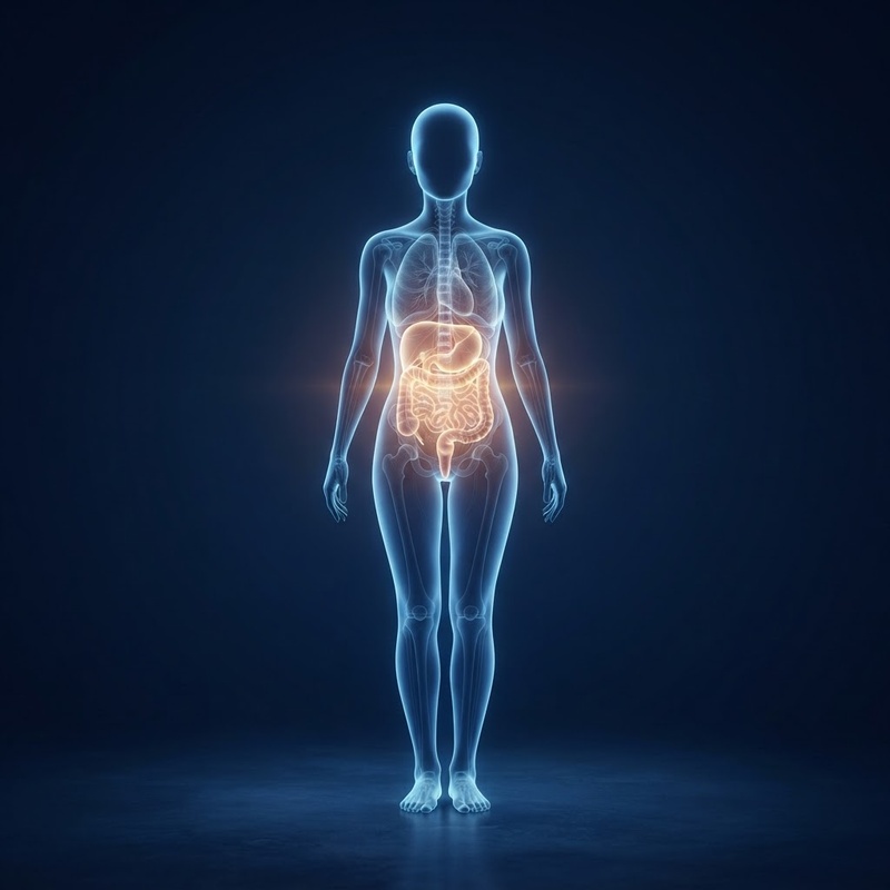
For many years, body fat was viewed simply as an inert storage depot for excess calories. Modern endocrinology and metabolic research have completely rewritten this picture. Today, adipose tissue is recognized as a complex, dynamic endocrine organ that actively communicates with the brain, liver, muscles, and immune system through biochemical signals.
For many years, body fat was viewed simply as an inert storage depot for excess calories. Modern endocrinology and metabolic research have completely rewritten this picture. Today, adipose tissue is recognized as a complex, dynamic endocrine organ that actively communicates with the brain, liver, muscles, and immune system through biochemical signals.
Crucially, where fat is deposited throughout the body plays a major role in how it interacts with human physiology. The two primary types of fat stored in the human body—subcutaneous adipose tissue and visceral adipose tissue—possess fundamental differences in anatomical placement, cellular architecture, hormonal sensitivity, and metabolic function. Understanding these distinctions provides valuable insight into how body composition influences overall metabolic health and energy regulation.
Understanding the Anatomical and Physiological Differences
To grasp why these two fat depots exert distinct metabolic effects, it is helpful to start with their physical distribution and anatomical orientation within the body.
Subcutaneous Fat: The Surface Layer
Subcutaneous adipose tissue (SAT) resides directly beneath the skin layer, lying above the abdominal wall and muscular framework. It accounts for approximately 85% to 90% of total body fat in most individuals. Subcutaneous fat is the tissue you can physically pinch on your arms, thighs, hips, and abdomen.
Physiologically, subcutaneous fat performs essential baseline functions. It acts as an insulator against thermal heat loss, provides mechanical cushioning to protect muscles and bones, and serves as a long-term energy reservoir. Under healthy metabolic conditions, subcutaneous tissue readily expands to store surplus circulating lipids, safely sequestering fat away from vital organs.
Visceral Fat: Deep Intra-Abdominal Storage
Visceral adipose tissue (VAT), in contrast, resides deep within the abdominal cavity. Rather than sitting beneath the skin, visceral fat is packed tightly around internal organs, including the liver, pancreas, stomach, intestines, and kidneys. Because it lies beneath the firm abdominal wall muscle layer, high levels of visceral fat can cause the abdomen to project outward into a firm, rounded shape, even when pinchable subcutaneous fat appears minimal.
Visceral fat typically accounts for roughly 10% to 15% of total body fat, but its metabolic influence is substantial. Because of its location deep inside the peritoneal space, visceral fat is closely integrated with the autonomic nervous system and the primary blood vessels servicing the digestive tract.
The Portal Vein Pathway
One of the most critical anatomical features distinguishing visceral fat from subcutaneous fat is its venous drainage route. Subcutaneous fat drains its metabolic products into systemic venous circulation, sending molecules through peripheral blood vessels.
Visceral fat, however, drains directly into the hepatic portal vein, which flows straight into the liver. This anatomical arrangement gives rise to what metabolic researchers refer to as the "portal hypothesis." When visceral fat breaks down lipid stores, it releases free fatty acids (FFAs) and signaling proteins directly into the portal circulation, exposing the liver to concentrated doses of metabolic fuels and inflammatory signals before these substances enter general circulation.
Endocrine Activity: Fat as an Active Metabolic Organ
Far from being passive energy tissue, both subcutaneous and visceral adipose tissues secrete biologically active signaling molecules known as adipokines. However, the endocrine profiles of visceral fat and subcutaneous fat are markedly different.
Receptor Sensitivity and Lipolytic Rate
Visceral adipose tissue is structurally distinct at the cellular level. Visceral fat cells (adipocytes) exhibit a higher density of beta-adrenergic receptors, which respond to catecholamines like adrenaline and noradrenaline by stimulating fat breakdown (lipolysis). At the same time, visceral fat cells have fewer alpha-2 adrenergic receptors, which normally inhibit lipolysis.
As a result, visceral fat has a higher baseline rate of lipolysis compared to subcutaneous fat, constantly mobilizing free fatty acids into portal blood. Subcutaneous fat cells are more stable and less prone to rapid lipolytic surges, making them better suited for steady, long-term energy storage.
Hormonal Signals: Leptin vs. Adiponectin
The two fat depots also differ in how they secrete key metabolic hormones:
- Leptin: Leptin communicates body energy reserves to the hypothalamus, helping regulate appetite and energy balance. Subcutaneous fat produces higher relative amounts of leptin than visceral fat. In healthy states, circulating leptin signals to the brain that energy stores are sufficient.
- Adiponectin: Adiponectin is a protective adipokine that enhances insulin sensitivity and exerts anti-inflammatory effects across vascular tissue. Both fat types secrete adiponectin, but expanding visceral fat tissue tends to suppress its secretion. Research indicates that elevated visceral fat levels correlate with lower circulating adiponectin concentrations, which can affect cellular glucose responsiveness.
Inflammatory Signaling and Cytokine Production
Beyond baseline hormone secretion, the degree of immune cell activation within adipose tissue differs sharply between subcutaneous and visceral depots.
Immune Cell Infiltration and Macrophage Activation
When fat tissue expands rapidly, individual fat cells can become enlarged (hypertrophic), experiencing cellular stress and localized hypoxia. In response, the tissue secretes chemoattractant signals that recruit immune cells, particularly macrophages, into the fat depot.
Visceral fat is far more susceptible to macrophage infiltration than subcutaneous fat. Furthermore, macrophages within visceral fat are more likely to adopt a pro-inflammatory "M1" activation state, forming microscopic clusters around stressed adipocytes that continuously produce inflammatory cytokines.
Pro-Inflammatory Molecules: IL-6 and TNF-Alpha
Visceral adipose tissue produces higher quantities of pro-inflammatory signaling proteins than subcutaneous fat, including:
- Interleukin-6 (IL-6): Up to one-third of circulating IL-6 originates from adipose tissue, with visceral fat contributing a disproportionate share. IL-6 travels via the portal vein to the liver, where it triggers production of C-reactive protein (CRP), a marker of systemic inflammation.
- Tumor Necrosis Factor-Alpha (TNF-alpha): TNF-alpha is a potent local and systemic inflammatory cytokine. Elevated TNF-alpha levels within visceral fat interfere with normal insulin receptor signaling inside adipocytes and adjacent tissues.
- Plasminogen Activator Inhibitor-1 (PAI-1): Visceral fat secretes elevated levels of PAI-1, a protein involved in regulating blood clotting dynamics and vascular signaling pathways.
This persistent inflammatory output from visceral fat creates an environment that can alter systemic metabolic signals throughout the body, contrasting with the relatively quiet profile of healthy subcutaneous fat.
Metabolic Impact: Glucose, Insulin, and Lipid Dynamics
Because visceral fat drains directly into the liver and secretes pro-inflammatory cytokines, it plays a prominent role in influencing systemic glucose regulation and lipid metabolism.
Hepatic Glucose Production and Insulin Dynamics
When the liver receives high concentrations of free fatty acids from visceral fat via the portal vein, its internal signaling adapts. The elevated influx of fatty acids promotes hepatic gluconeogenesis—the process by which the liver creates new glucose and releases it into the bloodstream. Concurrently, high fatty acid delivery can interfere with the liver's ability to clear insulin from circulation, altering systemic insulin levels.
Additionally, localized inflammatory cytokines like TNF-alpha disrupt insulin receptor substrate signaling inside liver cells. This makes hepatic tissue less responsive to insulin's signal to halt glucose release, contributing to higher fasting blood glucose patterns.
Peripheral Insulin Responsiveness and Lipid Shifts
The metabolic effects extend beyond the liver. Elevated circulating free fatty acids released from visceral fat can accumulate inside skeletal muscle cells—a phenomenon known as ectopic lipid deposition. Within muscle tissue, intracellular lipid metabolites interfere with glucose transporters, reducing muscle cells' ability to clear glucose from the blood in response to insulin.
Visceral fat storage also exerts a pronounced effect on circulating lipid profiles. Continuous delivery of free fatty acids to the liver stimulates overproduction of Very Low-Density Lipoproteins (VLDL), which leads to:
- Elevated serum triglyceride levels.
- A higher proportion of small, dense LDL particles.
- Reduced levels of High-Density Lipoprotein (HDL) cholesterol.
This distinct lipid pattern is closely linked with high visceral fat accumulation, whereas subcutaneous fat storage is generally not associated with these specific lipid shifts.
Lifestyle Strategies That Target Visceral Fat Accumulation
The physiological features that make visceral fat metabolically active—such as its high blood flow and abundance of catecholamine receptors—also make it responsive to positive lifestyle adjustments. Research indicates that during balanced lifestyle changes, visceral fat is often mobilized earlier than subcutaneous fat.
1. Aerobic Exercise and Movement
Physical activity is one of the most effective tools for encouraging visceral fat mobilization. Because visceral fat cells are sensitive to adrenaline and noradrenaline released during exercise, aerobic conditioning rapidly stimulates visceral fat breakdown.
- Moderate-to-Vigorous Aerobic Exercise: Activities such as brisk walking, jogging, cycling, or swimming for 150 to 300 minutes per week have been shown in studies to reduce visceral fat, even in the absence of major scale weight changes.
- Interval Training: High-Intensity Interval Training (HIIT) creates acute adrenergic surges that promote efficient lipolysis in intra-abdominal fat stores.
- Resistance Exercise: While aerobic exercise directly burns fuel, resistance exercise builds lean muscle tissue, which supports baseline insulin sensitivity and metabolic rate.
2. Dietary Adjustments for Metabolic Balance
Nutrition plays a central role in guiding fat storage patterns. Rather than restrictive severe diets, metabolic research emphasizes dietary quality and nutrient composition:
- Prioritizing Soluble Dietary Fiber: Soluble fiber (found in oats, legumes, flaxseeds, and vegetables) ferments in the gut to produce short-chain fatty acids (SCFAs). SCFAs help regulate satiety signals and support healthy liver lipid management.
- Reducing Refined Sugars and Fructose: Excess dietary fructose, particularly from sugar-sweetened beverages and highly processed foods, goes straight to the liver, where it can stimulate de novo lipogenesis (new fat creation).
- Focusing on Whole Foods: Emphasizing whole protein sources, healthy unsaturated fats, and complex carbohydrates stabilizes blood glucose spikes, minimizing insulin surges that promote fat storage.
3. Stress Regulation and Sleep Hygiene
Metabolic health extends beyond diet and exercise. Chronic psychological stress and inadequate sleep exert a direct influence on where the body stores fat.
- Cortisol Dynamics: Chronic stress maintains elevated levels of the stress hormone cortisol. Visceral fat tissue contains a higher density of glucocorticoid receptors than subcutaneous fat. When cortisol remains elevated alongside insulin, it signals the body to preferentially deposit energy as visceral fat inside the abdomen.
- Sleep Architecture: Sleep restriction disrupts appetite-regulating hormones, elevating ghrelin and reducing leptin. Studies indicate that insufficient sleep is associated with increased abdominal and visceral fat deposition.
Navigating Your Personal Metabolic Health Journey
Understanding the distinction between visceral fat and subcutaneous fat underscores an important truth in modern wellness: weight alone does not tell the full story of metabolic health. Two individuals with the exact same Body Mass Index (BMI) can have vastly different proportion ratios of visceral to subcutaneous fat, leading to distinct metabolic profiles.
Assessing body composition through waist-to-hip measurements, waist circumference tracking, bioelectrical impedance analysis, or DEXA scans can provide clearer insight into how fat is distributed in your body. Evaluating underlying metabolic markers—such as fasting glucose, fasting insulin, lipid panels, and inflammatory markers—offers the most meaningful picture of your health status.
Because individual genetics, hormonal patterns, age, and baseline health conditions vary widely, there is no single universal template for metabolic optimization. Partnering with a qualified healthcare professional allows you to explore personalized health assessments, interpret relevant lab markers accurately, and establish sustainable, evidence-based lifestyle habits tailored to your body.
If you are looking for clinical guidance tailored to your unique metabolic profile, explore EOS Health's personalized weight loss programs to connect with a licensed healthcare provider who can evaluate your health goals and design a comprehensive plan suited to your needs.
Glossary of Terms
Visceral Adipose Tissue (VAT)
Fat stored deep within the abdominal cavity that surrounds internal organs such as the liver, pancreas, and intestines, playing an active role in endocrine signaling and inflammation.
Subcutaneous Adipose Tissue (SAT)
Fat located directly underneath the skin layer that provides thermal insulation, energy storage, and physical cushioning for muscles and bones.
Hepatic Portal Vein
A major blood vessel that carries nutrient-rich blood and metabolic signaling molecules directly from the gastrointestinal tract and visceral fat tissue to the liver.
Adipokines
Cell-signaling proteins secreted by fat tissue (such as leptin and adiponectin) that help regulate appetite, inflammation, insulin sensitivity, and energy balance.
Free Fatty Acids (FFAs)
Molecules released during the breakdown of stored triglycerides that circulate in the bloodstream to be used as cellular energy or processed by the liver.
Insulin Resistance
A physiological state in which body tissues (muscle, liver, and fat) become less responsive to the hormone insulin, requiring higher levels of insulin to manage blood glucose levels.
Sources
faq
Common questions, answered.
What is the primary difference between visceral fat and subcutaneous fat?
Subcutaneous fat lies directly beneath the skin and serves as insulation and mechanical padding. Visceral fat is located deep inside the abdominal cavity, wrapping around internal organs like the liver and intestines. Visceral fat is more metabolically active and secretes higher levels of inflammatory signals into the portal blood system.
Why is visceral fat considered more harmful to metabolic health than subcutaneous fat?
Visceral fat drains directly into the hepatic portal vein, delivering free fatty acids and inflammatory cytokines directly to the liver. This can disrupt liver glucose management, impair insulin sensitivity, and alter circulating lipid levels far more than subcutaneous fat, which drains into general peripheral circulation.
Can you accurately measure visceral fat at home?
While definitive measurement requires imaging techniques like DEXA, CT, or MRI scans, simple home metrics provide useful estimates. Measuring waist circumference at the level of the navel or calculating your waist-to-hip ratio offers a practical proxy for tracking changes in abdominal fat distribution over time.
Does exercise target visceral fat specifically?
While spot reduction of fat is not possible, studies show that visceral fat is often mobilized earlier than subcutaneous fat during aerobic and high-intensity exercise. This occurs because visceral fat cells have a high density of adrenergic receptors that respond rapidly to catecholamines released during physical activity.
How does chronic stress contribute to visceral fat accumulation?
Chronic stress triggers sustained release of cortisol, a corticosteroid hormone. Visceral fat tissue has a high concentration of glucocorticoid receptors. When cortisol levels remain elevated alongside insulin, the body is signaled to preferentially deposit excess energy as intra-abdominal visceral fat.
Does dietary sugar intake affect visceral fat storage?
Yes, research indicates that high intake of refined sugars, particularly beverages rich in fructose, promotes liver fat accumulation and visceral fat storage. Fructose is processed primarily in the liver, where surplus amounts drive de novo lipogenesis (fat synthesis).
Is subcutaneous fat completely harmless?
Subcutaneous fat in normal amounts is essential for healthy insulation, cushioning, and endocrine regulation. However, when subcutaneous fat capacity is exceeded due to chronic energy surplus, extra lipids spill over into visceral depots and organ tissues, which can impact overall metabolic health.
How quickly can changes in visceral fat occur with lifestyle modifications?
Because visceral fat is highly sensitive to metabolic signals, improvements in dietary quality, regular aerobic exercise, and improved sleep quality can lead to reductions in visceral fat within weeks to months, even before significant changes in total body weight are visible.
Ready to feel like yourself again.™
Start free consultationwhat are you waiting for?
You're one click away from a new you.
Fill out our brief form and a licensed physician will evaluate which treatment plan is best for you.