Melasma vs. Sun Spots: How to Tell the Difference
Learn how to tell melasma vs. sun spots apart, exploring facial symmetry, hormonal triggers, and Wood's lamp evaluation to support your skin care journey.
Navigating changes in your skin can feel like an intricate puzzle, especially when unexpected dark patches or small brown spots appear on your face, neck, or chest. For many adult women, hyperpigmentation is a common experience influenced by genetics, sun exposure, and shifting hormonal landscapes. However, not all hyperpigmentation is created equal.
Navigating changes in your skin can feel like an intricate puzzle, especially when unexpected dark patches or small brown spots appear on your face, neck, or chest. For many adult women, hyperpigmentation is a common experience influenced by genetics, sun exposure, and shifting hormonal landscapes. However, not all hyperpigmentation is created equal. Two of the most widespread forms of facial discoloration—melasma and sun spots (solar lentigines)—frequently get mistaken for one another. While they may look somewhat similar at a glance, their underlying biological causes, patterns, and management approaches differ significantly.
Understanding the distinction between melasma and sun spots is far more than an academic exercise. Because the biological drivers behind these two conditions are distinct, attempting to address melasma with a routine designed solely for basic sun damage can sometimes lead to frustration or temporary pigment darkening. Conversely, identifying the true nature of your skin's pigment patterns allows you and your healthcare provider to develop an informed, gentle, and highly tailored skin care strategy. In this guide, we explore the science of melanogenesis, compare symmetry and color depth, discuss hormonal triggers, explain clinical Wood's lamp evaluations, and outline evidence-based approaches for supporting a balanced, healthy-looking complexion.
Understanding the Biological Mechanisms of Hyperpigmentation
To appreciate how melasma and sun spots differ, it helps to start at the cellular level with melanocytes—the pigment-producing cells located in the basal layer of your epidermis. Melanocytes produce melanin, the natural pigment responsible for skin tone. Melanin synthesis, known as melanogenesis, is a vital protective mechanism designed to absorb ultraviolet radiation and protect cellular DNA. However, when melanocytes become hyperactive, they produce excess melanin, which settles into surrounding keratinocytes and becomes visible as hyperpigmentation.
Solar Lentigines: Focused Photodamage in the Epidermis
Sun spots, known scientifically as solar lentigines or age spots, are discrete macules that develop primarily as a direct response to cumulative ultraviolet (UV) radiation over time. When your skin experiences years of sunlight exposure, local focal points of melanocytes experience chronic activation. This localized overproduction results in defined, flat brown spots that typically remain confined to the epidermal layer of the skin. Because solar lentigines stem directly from UV exposure, they appear predominantly on areas catching the most sun—such as cheekbones, nose bridge, forearms, hands, and upper chest.
Melasma: Hormonal Sensitivity and Microenvironmental Signals
Melasma involves a more dynamic and systemic underlying mechanism. While sun exposure acts as an essential trigger, the primary underlying driver is an increased sensitivity of melanocytes to hormonal fluctuations, particularly estrogen and progesterone. In melasma, melanocytes exhibit hyper-reactivity to multiple stimuli, including ultraviolet A (UVA), ultraviolet B (UVB), high-energy visible light (HEV or blue light), and heat. Furthermore, melasma involves microenvironmental factors, including localized vascular proliferation and basement membrane disruption, allowing melanin pigment to drop down into the deeper dermal layer. This key biological difference—epidermal versus mixed dermal-epidermal pigment deposition—explains why melasma requires a distinctly nuanced approach compared to standalone sun spots.
Key Visual Differences: Symmetry, Color Depth, and Distribution
When assessing dark spots on your face, careful observation of their pattern, boundaries, and coloration offers valuable insights. While only a licensed clinician can provide a formal evaluation, recognizing these visual characteristics fosters productive conversations during medical consultations.
Pattern and Bilateral Symmetry
One of the most characteristic features of melasma is its tendency toward bilateral symmetry across facial zones. Clinicians often categorize melasma into three distinct distribution patterns:
- Centrofacial pattern: Affecting the forehead, upper lip, chin, nose bridge, and central cheeks.
- Malar pattern: Focused on the prominent upper cheekbones and nose bridge.
- Mandibular pattern: Located along the jawline and chin.
If dark patches appear on your right cheek in a shape mirroring patches on your left cheek, or if you notice a characteristic "mustache-like" shadow along your upper lip, these patterns strongly align with melasma. Sun spots, by contrast, rarely exhibit bilateral symmetry. Instead, solar lentigines appear as individual, asymmetric, isolated spots scattered across areas receiving high UV radiation.
Edge Boundaries and Color Depth Variations
Examining the edges and color variations of pigmented areas provides another key clue:
- Sun spots (Solar Lentigines): Possess well-defined, sharp borders that can be easily outlined. Their color is usually uniform throughout, ranging from light tan to dark brown.
- Melasma: Features irregular, feathered, or soft borders merging gradually into surrounding skin. Color depth varies depending on pigment depth: epidermal melasma appears light to medium brown, whereas dermal melasma exhibits a grayish, slate-blue, or muddy hue due to light scattering through deeper tissue. Mixed melasma displays a combination of both brown and grayish tones.
Hormonal Triggers vs. Cumulative UV Radiative Stress
To understand why melasma behaves so differently from solar lentigines, it is essential to examine the physiological triggers behind each condition. While both respond to sunlight, their internal signaling pathways differ considerably.
The Hormonal Cascade in Melasma
Melasma is intricately tied to endocrine activity, particularly variations in female sex hormones. Estrogen and progesterone receptors exist on dermal melanocytes. When hormone levels fluctuate—such as during pregnancy, oral contraceptive use, hormone replacement therapy (HRT), or natural perimenopausal transitions—these hormones bind to melanocyte receptors, stimulating tyrosinase activity (the enzyme responsible for melanin synthesis).
Additionally, thyroid fluctuations and stress hormones like cortisol can influence melanocyte reactivity. Because melanocytes in melasma-prone skin remain in a state of heightened sensitivity, exposure to heat (such as saunas or hot cooking) or visible light can trigger pigment synthesis even when traditional UV sunlight is avoided.
Cumulative UV and Oxidative Stress in Sun Spots
Sun spots do not require a hormonal catalyst. Instead, they represent the cumulative historical record of ultraviolet radiation on skin cells. Over years of outdoor exposure, UV rays generate reactive oxygen species (ROS) and cellular DNA damage within epidermal keratinocytes. Keratinocytes continuously signal neighboring melanocytes to produce excess melanin. Over time, localized clusters of melanocytes remain permanently up-regulated, forming persistent solar lentigines that do not fluctuate with monthly hormonal cycles.
Clinical Evaluation Tools: Wood's Lamp and Diagnostic Care
Determining whether pigment is epidermal, dermal, or mixed is a critical step in guiding care. Licensed healthcare providers utilize specialized diagnostic tools during clinical evaluations to assess pigmentation accurately without relying solely on visual inspection.
Wood's Lamp Examination
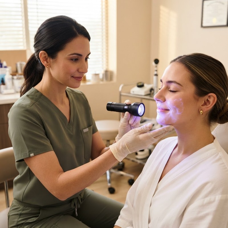
A classic diagnostic tool in clinical dermatology is the Wood's lamp—a specialized device emitting long-wave ultraviolet A (UVA) light around 365 nanometers. When shined on the skin in a dark room, Wood's light interacts differently with melanin depending on tissue depth:
- Epidermal pigment: Melanin located in the epidermis absorbs Wood's light strongly, creating sharp contrast and making dark areas appear more pronounced under illumination.
- Dermal pigment: Deep dermal melanin does not absorb light with sharp contrast; thus, dermal pigment does not accentuate under Wood's light and may appear muted.
- Mixed pigment: Certain areas accentuate under the lamp while other regions remain unchanged, confirming pigment across both epidermal and dermal layers.
Identifying pigment depth via Wood's lamp examination is vital. Epidermal pigment generally responds more readily to topical regimens, whereas dermal pigment requires a patient, multi-modal strategy overseen by a licensed provider.
Dermoscopy and Professional Diagnostics
In addition to Wood's lamp evaluation, clinicians use dermoscopy—high-powered magnification with polarized lighting—to examine pigment distribution patterns and vascular structures. Dermoscopy helps clinicians differentiate benign solar lentigines and melasma from atypical pigmented lesions, ensuring serious conditions are properly evaluated. Attempting to self-diagnose facial spots without professional guidance carries the risk of misidentifying lesions requiring direct medical attention.
Why Correct Identification Changes Your Skincare Strategy
Understanding whether you are addressing solar lentigines or melasma completely shifts how a clinician approaches your skin care plan. Because melasma involves hyper-reactive melanocytes and vascular components, aggressive interventions that work for sun spots can sometimes backfire if applied to melasma.
Caution with Heat and Aggressive Therapies
For standalone sun spots, localized physical modalities such as targeted cryotherapy, intense pulsed light (IPL), or Q-switched lasers are commonly used in clinical settings to disrupt localized epidermal pigment clusters. However, in individuals with melasma, high-heat energy from certain lasers or aggressive chemical peels can trigger post-inflammatory hyperpigmentation (PIH). Heat signals hyper-sensitive melanocytes to synthesize even more melanin, causing melasma patches to darken after temporary clearing. Therefore, melasma care prioritizes gentle, consistent topical management and inflammation control rather than high-intensity thermal treatments.
Comprehensive Sun and Light Protection
While broad-spectrum sunscreens protecting against UVA and UVB rays are essential for preventing solar lentigines, melasma care requires going further. Research shows that visible blue light (HEV light) penetrates deeply into the dermis and induces long-lasting hyperpigmentation in melasma-prone skin. To counteract this, dermatologists recommend broad-spectrum mineral sunscreens containing zinc oxide combined with iron oxides. Iron oxides act as a physical barrier reflecting visible light, providing daily protection against ambient indoor lighting, digital screens, and direct sunlight.
Evidence-Based Topical and Prescription Ingredients
When creating a personalized skin health strategy, a licensed healthcare provider may discuss various prescription and topical options tailored to your specific pigment depth and medical background:
- Tretinoin: Tretinoin is an FDA-approved prescription medication for acne, and it is commonly prescribed by clinicians for visible skin-aging signs. Retinoids support cell turnover and encourage the normal shedding of pigmented keratinocytes.
- Hydroquinone: Hydroquinone is a prescription-only ingredient in the United States that acts as a tyrosinase inhibitor to suppress excess melanin production. Due to its strength, hydroquinone is managed under close clinical supervision in structured cycle periods.
- Spironolactone: Spironolactone is an FDA-approved prescription medication as a diuretic, and it is commonly prescribed off-label for hormonal acne in women. While not a direct pigment treatment, managing hormonal acne flares reduces secondary post-inflammatory hyperpigmentation.
- Compounded Custom Formulations: Clinicians may consider customized compounded options to address multi-factorial pigment pathways simultaneously. Compounded products—such as hydroquinone, tranexamic acid, and azelaic acid custom blends, or tretinoin and clindamycin custom blends, as well as GHK-Cu copper peptide cream—are not FDA-approved, but offer tailored prescription combinations formulated by compounding pharmacies upon a licensed provider's order.
These prescription ingredients require an evaluation by a qualified dermatologist or telehealth clinician to determine if they are appropriate for your individual medical profile and skin goals.
Partnering with EOS Health for Tailored Skin Care
Managing facial hyperpigmentation—whether melasma, sun spots, or a combination of both—requires patience, scientific precision, and a supportive medical team. Because skin biology varies widely from person to person, there is no one-size-fits-all solution. Attempting to navigate over-the-counter options without understanding your pigment depth can lead to unnecessary expense and skin irritation.
At EOS Health, our clinical team provides comprehensive telehealth evaluations designed to make specialized care accessible from home. By evaluating your unique pattern, lifestyle factors, and hormonal history, our licensed medical providers can help clarify your skin concerns and design an individualized care plan tailored to support your long-term skin health.
If you are ready to explore clinician-guided options and learn more about personalized prescription formulas designed to support healthy aging and even skin tone, visit EOS Health Skincare Services today to start your consultation.
Melanogenesis
The complex biochemical process by which specialized epidermal cells called melanocytes produce melanin pigment in response to ultraviolet radiation, hormonal signals, or inflammation.
Solar Lentigines
Benign, flat brown macules (commonly called sun spots or age spots) caused by localized overproduction of melanin in response to chronic cumulative ultraviolet exposure.
Wood's Lamp Examination
A diagnostic technique utilizing a long-wave ultraviolet light (365 nm) to assess the depth of skin pigmentation and differentiate between epidermal, dermal, and mixed pigment deposition.
Compounded Medication
A customized medication prepared by a licensed pharmacist upon a physician's prescription to combine specific ingredients or tailor dosages; compounded products are not FDA-approved.
High-Energy Visible (HEV) Light
Short-wavelength, high-frequency light in the blue-violet spectrum (400-500 nm) emitted by sunlight and electronic screens, which can stimulate melanin synthesis in melasma-prone skin.
Sources
faq
Common questions, answered.
Can sun spots turn into melasma over time?
No, sun spots do not turn into melasma. Solar lentigines and melasma stem from different biological mechanisms. Sun spots result from localized epidermal photodamage caused by cumulative UV exposure, whereas melasma is driven by hormonal sensitivities combined with light and heat triggers. However, it is common to have both sun spots and melasma on your face at the same time.
How does a clinician determine if my melasma is epidermal or dermal?
A healthcare provider typically uses a Wood's lamp (a specialized 365nm UVA light source) during a clinical examination. Under Wood's light, epidermal melasma becomes more sharply defined and appears darker, while dermal melasma shows minimal contrast accentuation due to the deeper placement of pigment in the dermal layer.
Why is hydroquinone available only by prescription in the US?
Hydroquinone is a prescription-only ingredient in the United States to ensure safe usage under licensed medical supervision. Because improper long-term use can lead to adverse effects such as exogenous ochronosis or irritation, clinicians manage hydroquinone in carefully structured treatment cycles.
Are compounded skincare products FDA-approved?
No, compounded products—such as hydroquinone, tranexamic acid, and azelaic acid custom blends, or tretinoin and clindamycin custom blends, as well as GHK-Cu copper peptide cream—are not FDA-approved. Compounded medications are custom-mixed by licensed compounding pharmacies based on an individual prescription from a licensed healthcare provider.
What is the FDA approval status of tretinoin and spironolactone?
Tretinoin is an FDA-approved prescription medication for acne, and it is commonly prescribed by clinicians for visible skin-aging signs. Spironolactone is an FDA-approved prescription medication as a diuretic, and it is commonly prescribed off-label for hormonal acne in women.
Can heat alone trigger melasma flare-ups?
Yes. Melanocytes in melasma-prone skin are hyper-sensitive to thermal heat as well as light. Activities that raise skin temperature—such as hot saunas, hot yoga, steam rooms, or prolonged cooking over high heat—can induce localized cutaneous vasodilation and inflammatory signals that stimulate melanin synthesis, even in the absence of direct sunlight.
Why do mineral sunscreens with iron oxides work better for melasma?
Standard sunscreens block UVA and UVB rays, but high-energy visible (HEV) blue light from the sun and digital screens can also trigger melasma pigment. Mineral sunscreens formulated with zinc oxide and iron oxides provide a physical barrier that reflects visible light wavelengths, helping protect melasma-prone skin from blue light stimulation.
Can melasma improve during or after pregnancy?
Melasma that develops during pregnancy (often called chloasma) is linked to elevated estrogen and progesterone levels. For some women, pigment intensity may gradually soften months after delivery as hormone levels stabilize, though persistent cases benefit from clinical evaluation by a healthcare provider.
Great skin starts with the right protocol.™
Start free consultationwhat are you waiting for?
You're one click away from a new you.
Fill out our brief form and a licensed physician will evaluate which treatment plan is best for you.