Why Autophagy Research Is Central to Healthy Longevity Science
Explore how autophagy research shapes healthy longevity science. Discover how cellular recycling supports healthy aging and metabolic wellness at EOS Health.
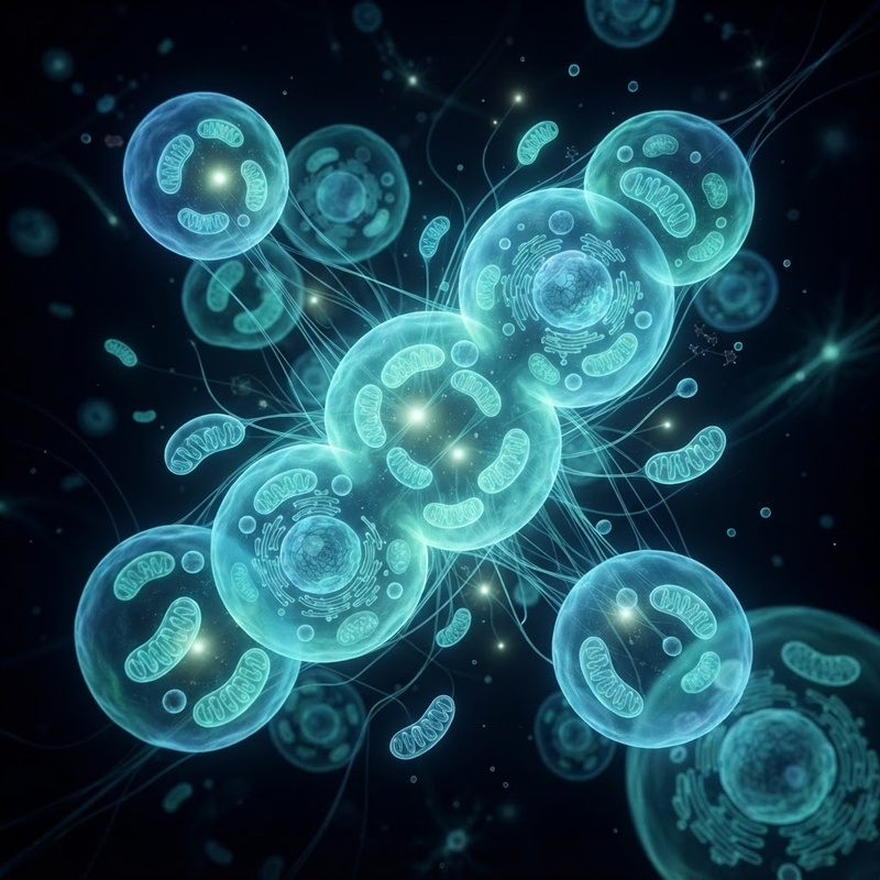
Every moment of the day, trillions of cells in the human body carry out complex metabolic processes necessary for vitality. Over time, normal cellular activity generates metabolic byproducts, worn-out organelles, and misfolded proteins. To maintain balance, energy, and physiological resilience, tissues depend on internal housekeeping systems that continually refresh cellular components. At the forefront of modern longevity science is autophagy—a fundamental cellular self-cleaning process that allows cells to break down and recycle their own damaged parts.
Every moment of the day, trillions of cells in the human body carry out complex metabolic processes necessary for vitality. Over time, normal cellular activity generates metabolic byproducts, worn-out organelles, and misfolded proteins. To maintain balance, energy, and physiological resilience, tissues depend on internal housekeeping systems that continually refresh cellular components. At the forefront of modern longevity science is autophagy—a fundamental cellular self-cleaning process that allows cells to break down and recycle their own damaged parts.
Derived from Greek roots meaning "self-eating," autophagy was once viewed simply as a stress response to nutrient deprivation. Today, clinical researchers recognize it as a highly regulated quality-control system essential for cellular maintenance and healthy physiological aging. As interest in healthy longevity science grows, understanding how autophagy functions—and how lifestyle choices may influence its signaling pathways—empowers individuals to make informed decisions about their health journeys. At EOS Health, we explore evidence-based approaches to metabolic wellness, supporting healthy aging through comprehensive clinical guidance and personalized care.
Understanding the Biology of Autophagy and Cellular Clean-Up
At its core, autophagy is the body's primary mechanism for intracellular recycling. Rather than allowing damaged cellular components to accumulate, cells wrap degraded material in double-membraned vesicles called autophagosomes. These structures then transport their contents to lysosomes—specialized organelles containing digestive enzymes—where the material is broken down into basic building blocks like amino acids, fatty acids, and sugars. These recycled molecules are subsequently released back into the cytoplasm to be reused for energy production or new protein synthesis. To visualize this process, imagine a municipal recycling center where damaged infrastructure is systematically dismantled and reprocessed into raw construction materials to maintain city stability.
Scientists classify autophagy into three distinct biological types based on how cellular material reaches the lysosome:
- Macroautophagy: The most widely studied form, wherein larger cellular structures, damaged organelles, and protein aggregates are sequestered within autophagosomes before lysosomal breakdown.
- Microautophagy: A direct process where the lysosomal membrane itself engulfs small portions of cytoplasmic material through direct invagination.
- Chaperone-Mediated Autophagy (CMA): A targeted pathway in which specialized chaperone proteins identify specific target molecules containing particular peptide sequences and deliver them directly into the lysosome.
At the molecular level, macroautophagy involves a coordinated cascade of protein complexes. When initiated, the ULK1 kinase complex signals the formation of a crescent-shaped membrane structure known as the phagophore. As this membrane expands to enclose cellular debris, a key protein called LC3-I undergoes lipidation to become LC3-II, inserting into the autophagosomal membrane. Once fully enclosed, SNARE proteins facilitate lysosomal fusion to degrade enclosed contents.
Regulation of autophagy relies heavily on key nutrient-sensing enzyme networks, primarily mTOR (mechanistic target of rapamycin) and AMPK (AMP-activated protein kinase). When nutrients—particularly amino acids and glucose—are abundant, mTOR activity increases, signaling the cell to prioritize growth and protein synthesis while dampening autophagy. Conversely, when cellular energy levels dip or stress rises, AMPK is activated, inhibiting mTOR and stimulating autophagy pathways. This delicate metabolic balance ensures that cells alternate between growth and maintenance according to physiological needs.
How Scientists Study Autophagy in Healthy Aging Research
One of the central questions in longevity science is why cellular function tends to decline over time. Research suggests that as organisms age, natural autophagic efficiency gradually declines. This reduction in housekeeping capacity can lead to the gradual accumulation of cellular debris, including misfolded proteins, lipofuscin pigments, and dysfunctional organelles that impair tissue function.
In particular, researchers pay close attention to mitophagy—the selective degradation and recycling of damaged mitochondria. Mitochondria generate cellular ATP, but damaged mitochondria produce elevated reactive oxygen species (ROS). The molecular pathway governing mitophagy relies on PINK1 and Parkin proteins. When a mitochondrion suffers membrane depolarization, PINK1 accumulates on its outer membrane, recruiting Parkin to tag the damaged organelle for autophagic sequestration. By clearing impaired mitochondria before they compromise tissue function, mitophagy helps maintain cellular energy balance and metabolic stability.
Emerging scientific literature also explores the connection between declining autophagic flux and cellular senescence. Senescent non-dividing cells secrete pro-inflammatory factors known as the senescence-associated secretory phenotype (SASP). Research indicates that robust autophagic processes help clear damaged cellular components before cells undergo senescent arrest. In laboratory models, observational enhancement of autophagic pathways correlates with improved cellular stress tolerance and preserved physiological markers across diverse organisms.
Human clinical studies continue to evaluate how autophagy markers change with age, physical activity, and dietary patterns. While measuring real-time autophagic flux in living human tissue presents technical challenges, researchers utilize indirect surrogate biomarkers. These include measuring LC3-II/LC3-I protein ratios in peripheral blood mononuclear cells, plasma metabolomic profiling, and tissue biopsies. Scientists emphasize that autophagy is an ongoing biological process requiring physiological balance: optimal cellular function relies on appropriate cycles of renewal rather than continuous, unmoderated activation.
Lifestyle Factors and Autophagy Induction: What the Evidence Shows
While pharmacological modulators of autophagy remain an active area of clinical investigation, researchers have identified several natural physiological stimuli that engage autophagic pathways. Lifestyle factors such as physical movement, dietary structure, and restorative sleep play integral roles in supporting cellular maintenance systems.
1. Physical Exercise and Movement
Physical activity stands out as one of the most potent non-pharmacological triggers of cellular clean-up. Studies indicate that both endurance cardiovascular exercise and resistance training induce autophagy in skeletal muscle, cardiac tissue, metabolic organs, and neural networks. During exercise, muscular contraction increases energy demand, elevating the intracellular AMP-to-ATP ratio. This metabolic shift activates AMPK, prompting cells to clear damaged components to sustain energy demands.
Furthermore, regular movement stimulates mitochondrial biogenesis alongside mitophagy via PGC-1α signaling networks. For instance, combining zone 2 cardiovascular training with periodic resistance sessions provides consistent metabolic stimuli that support cellular quality control, physical stamina, and systemic metabolic health as adults age.
2. Dietary Timing and Caloric Modulation
Nutrient availability directly signals cellular nutrient sensors. Research exploring caloric modulation and time-restricted feeding suggests that transient periods of nutrient scarcity lower circulating insulin and amino acid levels, thereby easing mTOR suppression of autophagy. During brief fasting intervals, cells transition from an anabolic storage mode to a catabolic maintenance mode, initiating cellular recycling pathways.
Mechanistically, after hepatic glycogen stores diminish—typically within 12 to 16 hours depending on activity levels and metabolic rate—cells rely increasingly on fatty acid oxidation and autophagic breakdown for substrate energy. However, scientists note that individual responses to fasting vary significantly depending on baseline metabolic health, physical activity, age, and nutritional status. Sustainable nutrition plans meeting essential macronutrient and micronutrient needs remain paramount, as extreme starvation can lead to muscle loss rather than physiological renewal.
3. Restorative Sleep and Circadian Integrity
Emerging research underscores the profound connection between circadian rhythms and cellular maintenance. Many biological processes, including brain-specific clearing systems and liver metabolic pathways, operate on rhythmic daily cycles coordinated by clock genes such as BMAL1 and CLOCK.
During deep slow-wave sleep, neural tissues utilize the glymphatic system to flush metabolic byproducts from the interstitial space of the brain. This central cleaning process works alongside localized neuronal autophagy to maintain metabolic balance. Quality sleep supports nocturnal cellular repair mechanisms, ensuring that tissues undergo structural maintenance during resting hours. Conversely, disruptions in sleep architecture or shift-work schedules can alter clock gene expression, potentially influencing daytime cellular resilience.
Dietary Factors and Caloric Mimetics Under Investigation
In addition to exercise and fasting research, scientific interest has focused on naturally occurring compounds that interact with autophagy pathways, often called "caloric restriction mimetics." Preclinical studies have evaluated several dietary phytochemicals for their potential to support cellular recycling signals:
- Spermidine: A naturally occurring polyamine found in wheat germ, aged cheese, mushrooms, and legumes that interacts with acetyltransferase enzymes (such as EP300) to support autophagic gene signaling.
- Resveratrol and Polyphenols: Plant compounds present in grapes, berries, and green tea (such as EGCG) that engage SIRT1 and AMPK pathways in laboratory models, supporting cellular stress responses.
- Quercetin and Fisetin: Flavonoids abundant in apples, onions, and leafy greens, studied for their roles in modulating cellular stress responses and supporting tissue balance.
- Sulforaphane: An isothiocyanate derived from cruciferous vegetables like broccoli sprouts, studied for its ability to activate Nrf2 signaling, coordinating antioxidant defenses with cellular clearing machinery.
While preclinical findings are promising, nutritional scientists emphasize that dietary compounds work best within a balanced lifestyle foundation. The bioavailability of polyphenol molecules varies among individuals due to gut microbiome composition and digestive absorption. No single compound or supplement acts as a substitute for foundational health habits like adequate protein intake, diverse nutrient consumption, regular physical movement, and professional clinical oversight. Furthermore, dietary supplements should be selected thoughtfully, as individual tolerability and metabolic needs differ.
Personalized Longevity Care: Navigating Science with Clinical Guidance
The science of autophagy illustrates a core principle of modern functional health: cellular wellness is a holistic, interconnected dynamic. Because metabolic rate, hormone levels, cardiovascular markers, and cellular stress profiles vary widely from person to person, there is no one-size-fits-all protocol for optimizing cellular health. Unsupervised implementation of extreme fasting regimens or high-dose supplement stacks can sometimes lead to unintended outcomes, such as muscle loss, electrolyte imbalances, or hormonal disruption.
Partnering with a licensed clinical team provides a safe, evidence-based structure for exploring longevity science. Comprehensive clinical evaluations utilize routine biomarker labs—including fasting insulin, HbA1c, lipid subfractions, inflammatory markers like high-sensitivity C-reactive protein (hs-CRP), and liver enzymes—to establish an accurate metabolic baseline. A qualified healthcare provider can evaluate metabolic flexibility and individual goals, crafting a personalized wellness plan that supports natural cellular maintenance safely and sustainably.
Supporting Your Cellular Health Journey with EOS Health
Healthy longevity is not about chasing quick fixes or unproven trends; it is about adopting evidence-informed habits that nurture cellular vitality over a lifetime. Understanding the science of autophagy empowers individuals to appreciate how movement, balanced nutrition, restorative rest, and personalized care work together to support the body's innate maintenance systems.
At EOS Health, our clinical team provides comprehensive, telehealth-based guidance designed around your personal health profile. Whether seeking to optimize metabolic health, evaluate key lab markers, or establish a sustainable lifestyle framework, our licensed clinicians are here to support your journey every step of the way. Explore our tailored programs today through EOS Health Longevity Care and discover how proactive medical guidance can help you thrive.
Glossary of Key Longevity Terms
Autophagy
A regulated biological process by which cells break down and recycle their own worn-out components, such as damaged proteins and organelles, to maintain cellular efficiency and balance.
Mitophagy
A targeted form of autophagy specifically responsible for identifying, clearing, and recycling damaged or dysfunctional mitochondria within cells.
mTOR (Mechanistic Target of Rapamycin)
A key nutrient-sensing enzyme complex that regulates cellular growth, protein synthesis, and metabolism in response to nutrient availability and growth factors.
AMPK (AMP-Activated Protein Kinase)
An essential cellular energy sensor that becomes activated when energy levels are low, stimulating energy-generating pathways and initiating autophagy.
Autophagosome
A double-membraned cellular vesicle that engulfs damaged proteins and organelles, transporting them to the lysosome for degradation and recycling.
Sources
faq
Common questions, answered.
What is autophagy in simple terms?
Autophagy is the body's natural cellular recycling system. It allows cells to identify, disassemble, and recycle old, damaged, or dysfunctional cellular components, converting them into energy and raw materials for cellular renewal.
How does fasting influence autophagy in human research?
Research indicates that fasting temporarily reduces nutrient availability, which decreases mTOR activity and activates AMPK signaling. This metabolic shift prompts cells to initiate cellular clean-up. However, the exact duration required to induce significant autophagy in humans varies based on individual metabolism, physical activity, and health status.
Can regular physical exercise activate cellular clean-up?
Yes. Physical exercise is one of the most effective ways to support autophagy in muscles, heart tissue, and metabolic organs. Exercise increases cellular energy demand, stimulating AMPK pathways that encourage mitochondrial recycling and cellular maintenance.
Are there dietary supplements that promote autophagy?
Preclinical studies have investigated several natural compounds, such as spermidine, resveratrol, and green tea polyphenols, for their potential to support autophagy signaling. While research is ongoing, these compounds are best viewed as complementary to a healthy lifestyle under clinical supervision rather than standalone solutions.
How do researchers measure autophagy in living tissues?
In research settings, scientists measure autophagy using specialized techniques such as tracking autophagosomes via electron microscopy, measuring specific protein markers like LC3-II and p62, or assessing metabolic flux in blood and tissue samples. Clinical measures in humans focus primarily on related metabolic biomarkers.
Is higher autophagy activity always beneficial?
Autophagy requires balance. While periodic activation supports cellular maintenance and stress resilience, excessive or uncontrolled autophagy can lead to unwanted cellular breakdown. The body functions best when cellular clean-up operates in harmony with cellular growth and protein synthesis.
How does autophagy relate to mitochondrial health?
A specialized form of autophagy called mitophagy specifically targets damaged or inefficient mitochondria. By clearing impaired mitochondria, mitophagy helps maintain clean energy production and reduces cellular oxidative stress.
Why is consulting a clinician recommended before starting fasting or longevity protocols?
Personalized health guidance ensures that lifestyle changes—such as fasting, intense exercise regimens, or dietary supplements—align with your specific medical history, nutritional status, and health goals. A licensed clinician can monitor vital health markers, address potential risks, and optimize your plan safely.
Invest in the years ahead.™
Start free consultationwhat are you waiting for?
You're one click away from a new you.
Fill out our brief form and a licensed physician will evaluate which treatment plan is best for you.